



Heezy1323
-
Posts
261 -
Joined
-
Last visited
-
Days Won
2
Content Type
Profiles
News
Minnesota Twins Videos
2026 Minnesota Twins Top Prospects Ranking
2022 Minnesota Twins Draft Picks
Minnesota Twins Free Agent & Trade Rumors, Notes, & Tidbits
Guides & Resources
2023 Minnesota Twins Draft Picks
The Minnesota Twins Players Project
2024 Minnesota Twins Draft Picks
2025 Minnesota Twins Draft Pick Tracker
2026 Minnesota Twins Draft Tracker: Picks & Bonuses
Forums
Blogs
Events
Store
Downloads
Gallery
Everything posted by Heezy1323
-
Royce Lewis: Patellar Tendinopathy
Heezy1323 replied to Heezy1323's topic in Twins Daily Front Page News
I think his age only contributes to the extent that for the most part only young people are active enough to get this problem. By age 19, most males are done growing, though it's possible he isn't. If he isn't, that could be a factor. I think the most likely issue is relative overuse, given that he has likely played more baseball games this year than he ever has previously. He likely just needs some time to figure out what it is going to take to keep his body in the best condition possible while playing professional sports. The answer to that is different for everyone, and often takes some experience to find the best methods for an individual. -
Royce Lewis: Patellar Tendinopathy
Heezy1323 replied to Heezy1323's topic in Twins Daily Front Page News
Thanks for the question. I wouldn't think so. Remember, this area is very small (around the size of a couple of peas), so we aren't talking about extensive involvement here. In this context, the term 'degenerative' refers more to the body's ability to rebuild this tissue in response to stress. When that balance is thrown out of whack by one or more factors, the tissue 'build-up' is unable to keep up with the tissue 'break-down' and the tissue starts to become 'degenerative'. Improving the balance of this equation can (and should) restore the tissue to a healthy condition. It may be something he needs to do maintenance exercises for going forward to prevent recurrence, but my hope would be that is the extent of it. -
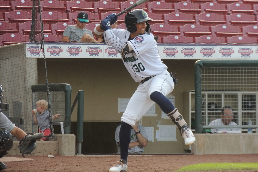
(Twins Daily Note: In a Jeff Johnson story in The Gazette from Cedar Rapids, Royce Lewis noted that he has been diagnosed with Patellar tendinitis and he has been playing with the injury for about a month. He did play in the Midwest League All Star game on Tuesday night, so it is considered a minor injury. Today, Heezy tells us all about patellar tendinitis.) The Twins community recently received some concerning news about highly-regarded prospect Royce Lewis. Lewis left the Cedar Rapids Kernels Saturday game with what is being reported as ‘patellar tendinitis’, which has been causing him trouble off-and-on for about a month. But what is ‘patellar tendinitis’ exactly? And what might it mean for Royce going forward? Let’s discuss: Image courtesy of Seth Stohs, Twins Daily (photo of Royce Lewis) (Twins Daily Note: In a Jeff Johnson story in The Gazette from Cedar Rapids, Royce Lewis noted that he has been diagnosed with Patellar tendinitis and he has been playing with the injury for about a month. He did play in the Midwest League All Star game on Tuesday night, so it is considered a minor injury. Today, Heezy tells us all about patellar tendinitis.) The Twins community recently received some concerning news about highly-regarded prospect Royce Lewis. Lewis left the Cedar Rapids Kernels Saturday game with what is being reported as ‘patellar tendinitis’, which has been causing him trouble off-and-on for about a month. But what is ‘patellar tendinitis’ exactly? And what might it mean for Royce going forward? Let’s discuss: Question 1: Where is the ‘patella tendon’? The patella tendon is the tendon that goes from your kneecap (also called the patella) down to the upper part of your shin bone (tibia). It is the attachment of your quadriceps muscle group to your lower leg, and it is what allows people to extend the knee. Question 2: What is ‘patellar tendinitis’? Patella tendinitis is a term commonly used to refer to activity-related pain that occurs near the attachment of the patella tendon to the patella. The suffix ‘-itis’ is used to indicate inflammation. While the term is commonly used, in actuality a more appropriate term is ‘patella tendinopathy’, which refers to degenerative changes within the tendon in the absence of inflammation (which is more accurately the case in this diagnosis). This difference is important when considering treatment options. Question 3: Royce is clearly a high-level athlete. Why did he get this problem? Patellar tendinopathy is also commonly called ‘jumper’s knee’ since it occurs most frequently in athletes that do a lot of jumping. In some studies of professional volleyball and basketball players, the incidence of jumper’s knee has been shown to be more than 30%. It is much less common in non-jumping athletes, but still occurs in around 2-3% of soccer players. I was not able to find any information specifically discussing the incidence in baseball players. It is unclear exactly why this problem occurs. It is most likely a combination of factors including BMI, flat feet, muscle imbalance in the quad/hamstrings, low flexibility, and intrinsic properties of the patellar tendon. There are likely other factors as well, including overuse. The area involved is usually located directly at the bottom end of the patella/top part of the tendon. Symptoms usually come on gradually over time. Initially, the knee typically hurts only with activity. Over time, if the condition worsens, pain may begin to be present even at rest. Question 4: How is patella tendinopathy diagnosed? The diagnosis is usually fairly clear from the history and physical exam of the athlete. Xrays are usually normal, though in some cases calcifications of the tendon may be visible. An MRI is the standard test to identify the extent of the problem and also to rule out other problems inside the knee. The area of the tendon involved in the problem is typically fairly small- around the size of a couple tic-tacs. Question 5: How is patella tendinopathy treated? The most commonly prescribed treatment for patellar tendinopathy is rest from vigorous activity and specific physical therapy exercises (called eccentric exercises). These exercises are designed to strengthen the quad muscles, stretch the hamstrings and ultimately cause favorable adaptation of the knee. The time needed for symptoms to resolve can be highly variable, but often takes at least a few weeks. When therapy isn’t effective, other treatments can be tried including various injections and ultrasound. At this time, there is no significant evidence that PRP (platelet rich plasma) injections are helpful for this condition, though I suspect it is being considered. There is, to my knowledge, no significant data on stem cell injections for this problem. Question 6: Is surgery ever needed for patellar tendinopathy? Rarely, yes. In most studies, around 10% of patients will fail to respond to appropriate conservative treatment. In these cases, surgery may be needed. There are two main options: open surgery and arthroscopic surgery. In either case, the procedure is similar- the area of affected tendon is excised and a small (a few millimeters) part of the patella bone is removed to stimulate healing. Therapy is begun soon after surgery. The success rate for return to sports is around 80% for both surgeries, with return after the arthroscopic version being quicker on average. Usually, 4-6 months is needed for full return to sports after surgery. Question 7: Is Royce at increased risk of rupturing the patella tendon because of this problem? No. Having patella tendinopathy does not appear to place anyone at increased risk of having a patella tendon rupture when compared to those without the problem. Overall, I believe the most likely scenario to be that Lewis’ body is adjusting to playing professional baseball every day and he is having some minor issues as a result. I don’t expect this to be a substantial problem going forward, though the possibility that this requires surgery in the future does exist. Hopefully he will get through rehab quickly and be back on the field soon. View full article
-
 Question 1: Where is the ‘patella tendon’? The patella tendon is the tendon that goes from your kneecap (also called the patella) down to the upper part of your shin bone (tibia). It is the attachment of your quadriceps muscle group to your lower leg, and it is what allows people to extend the knee. Question 2: What is ‘patellar tendinitis’? Patella tendinitis is a term commonly used to refer to activity-related pain that occurs near the attachment of the patella tendon to the patella. The suffix ‘-itis’ is used to indicate inflammation. While the term is commonly used, in actuality a more appropriate term is ‘patella tendinopathy’, which refers to degenerative changes within the tendon in the absence of inflammation (which is more accurately the case in this diagnosis). This difference is important when considering treatment options. Question 3: Royce is clearly a high-level athlete. Why did he get this problem? Patellar tendinopathy is also commonly called ‘jumper’s knee’ since it occurs most frequently in athletes that do a lot of jumping. In some studies of professional volleyball and basketball players, the incidence of jumper’s knee has been shown to be more than 30%. It is much less common in non-jumping athletes, but still occurs in around 2-3% of soccer players. I was not able to find any information specifically discussing the incidence in baseball players. It is unclear exactly why this problem occurs. It is most likely a combination of factors including BMI, flat feet, muscle imbalance in the quad/hamstrings, low flexibility, and intrinsic properties of the patellar tendon. There are likely other factors as well, including overuse. The area involved is usually located directly at the bottom end of the patella/top part of the tendon. Symptoms usually come on gradually over time. Initially, the knee typically hurts only with activity. Over time, if the condition worsens, pain may begin to be present even at rest. Question 4: How is patella tendinopathy diagnosed? The diagnosis is usually fairly clear from the history and physical exam of the athlete. Xrays are usually normal, though in some cases calcifications of the tendon may be visible. An MRI is the standard test to identify the extent of the problem and also to rule out other problems inside the knee. The area of the tendon involved in the problem is typically fairly small- around the size of a couple tic-tacs. Question 5: How is patella tendinopathy treated? The most commonly prescribed treatment for patellar tendinopathy is rest from vigorous activity and specific physical therapy exercises (called eccentric exercises). These exercises are designed to strengthen the quad muscles, stretch the hamstrings and ultimately cause favorable adaptation of the knee. The time needed for symptoms to resolve can be highly variable, but often takes at least a few weeks. When therapy isn’t effective, other treatments can be tried including various injections and ultrasound. At this time, there is no significant evidence that PRP (platelet rich plasma) injections are helpful for this condition, though I suspect it is being considered. There is, to my knowledge, no significant data on stem cell injections for this problem. Question 6: Is surgery ever needed for patellar tendinopathy? Rarely, yes. In most studies, around 10% of patients will fail to respond to appropriate conservative treatment. In these cases, surgery may be needed. There are two main options: open surgery and arthroscopic surgery. In either case, the procedure is similar- the area of affected tendon is excised and a small (a few millimeters) part of the patella bone is removed to stimulate healing. Therapy is begun soon after surgery. The success rate for return to sports is around 80% for both surgeries, with return after the arthroscopic version being quicker on average. Usually, 4-6 months is needed for full return to sports after surgery. Question 7: Is Royce at increased risk of rupturing the patella tendon because of this problem? No. Having patella tendinopathy does not appear to place anyone at increased risk of having a patella tendon rupture when compared to those without the problem. Overall, I believe the most likely scenario to be that Lewis’ body is adjusting to playing professional baseball every day and he is having some minor issues as a result. I don’t expect this to be a substantial problem going forward, though the possibility that this requires surgery in the future does exist. Hopefully he will get through rehab quickly and be back on the field soon.
-
Royce Lewis: Patellar Tendinopathy Heezy1323 The Twins community recently received some concerning news about highly-regarded prospect Royce Lewis. Lewis left the Cedar Rapids Kernels Saturday game with what is being reported as ‘patellar tendinitis’, which has been causing him trouble off-and-on for about a month. But what is ‘patellar tendinitis’ exactly? And what might it mean for Royce going forward? Let’s discuss: Question 1: Where is the ‘patella tendon’? The patella tendon is the tendon that goes from your kneecap (also called the patella) down to the upper part of your shin bone (tibia). It is the attachment of your quadriceps muscle group to your lower leg, and it is what allows people to extend the knee. Question 2: What is ‘patellar tendinitis’? Patella tendinitis is a term commonly used to refer to activity-related pain that occurs near the attachment of the patella tendon to the patella. The suffix ‘-itis’ is used to indicate inflammation. While the term is commonly used, in actuality a more appropriate term is ‘patella tendinopathy’, which refers to degenerative changes within the tendon in the absence of inflammation (which is more accurately the case in this diagnosis). This difference is important when considering treatment options. Question 3: Royce is clearly a high-level athlete. Why did he get this problem? Patellar tendinopathy is also commonly called ‘jumper’s knee’ since it occurs most frequently in athletes that do a lot of jumping. In some studies of professional volleyball and basketball players, the incidence of jumper’s knee has been shown to be more than 30%. It is much less common in non-jumping athletes, but still occurs in around 2-3% of soccer players. I was not able to find any information specifically discussing the incidence in baseball players. It is unclear exactly why this problem occurs. It is most likely a combination of factors including BMI, flat feet, muscle imbalance in the quad/hamstrings, low flexibility, and intrinsic properties of the patellar tendon. There are likely other factors as well, including overuse. The area involved is usually located directly at the bottom end of the patella/top part of the tendon. Symptoms usually come on gradually over time. Initially, the knee typically hurts only with activity. Over time, if the condition worsens, pain may begin to be present even at rest. Question 4: How is patella tendinopathy diagnosed? The diagnosis is usually fairly clear from the history and physical exam of the athlete. Xrays are usually normal, though in some cases calcifications of the tendon may be visible. An MRI is the standard test to identify the extent of the problem and also to rule out other problems inside the knee. The area of the tendon involved in the problem is typically fairly small- around the size of a couple tic-tacs. Question 5: How is patella tendinopathy treated? The most commonly prescribed treatment for patellar tendinopathy is rest from vigorous activity and specific physical therapy exercises (called eccentric exercises). These exercises are designed to strengthen the quad muscles, stretch the hamstrings and ultimately cause favorable adaptation of the knee. The time needed for symptoms to resolve can be highly variable, but often takes at least a few weeks. When therapy isn’t effective, other treatments can be tried including various injections and ultrasound. At this time, there is no significant evidence that PRP (platelet rich plasma) injections are helpful for this condition, though I suspect it is being considered. There is, to my knowledge, no significant data on stem cell injections for this problem. Question 6: Is surgery ever needed for patellar tendinopathy? Rarely, yes. In most studies, around 10% of patients will fail to respond to appropriate conservative treatment. In these cases, surgery may be needed. There are two main options: open surgery and arthroscopic surgery. In either case, the procedure is similar- the area of affected tendon is excised and a small (a few millimeters) part of the patella bone is removed to stimulate healing. Therapy is begun soon after surgery. The success rate for return to sports is around 80% for both surgeries, with return after the arthroscopic version being quicker on average. Usually, 4-6 months is needed for full return to sports after surgery. Question 7: Is Royce at increased risk of rupturing the patella tendon because of this problem? No. Having patella tendinopathy does not appear to place anyone at increased risk of having a patella tendon rupture when compared to those without the problem. Overall, I believe the most likely scenario to be that Lewis’ body is adjusting to playing professional baseball every day and he is having some minor issues as a result. I don’t expect this to be a substantial problem going forward, though the possibility that this requires surgery in the future does exist. Hopefully he will get through rehab quickly and be back on the field soon.
-
So this is a very complex question/issue- I could probably do a two hour talk about it. No one wants to be inundated with that much ortho info by me, so I'll do my best to summarize. As discussed in the article above, the meniscus is critical for normal function of many aspects of the knee. This includes stability, cushion and protection of the surface cartilage. Because of the importance of the meniscus, it is critical to try to preserve as much meniscus as possible, as often as possible in order to optimize the long-term function/health of the knee. For example, we know from studies that one of the biggest risk factors for ACL graft failure after reconstruction is having had part of your meniscus removed at the same time as reconstruction. We also know that the greater the amount of meniscus removed, the greater the risk that a person develops arthritis down the road. Recall, arthritis is thinning or damage to the gliding surface cartilage of the knee (which is different from the meniscus itself). Once damage to the surface cartilage occurs, we don't have a great way to repair or restore the cartilage to normal. (There are some procedures that we have as options, but they are rarely as good as normal cartilage.) Because of the lack of good options to treat arthritis, especially in young patients, we have become more aggressive over time at attempting to repair meniscus tears in order to prevent this problem. With regards to stability, think of the meniscus as a chock that you place behind the wheel of a trailer that is parked on a hill to prevent it from rolling away. The meniscus helps stabilize the knee in the same way. As a result, when you remove part of the meniscus during an ACL reconstruction, there is a smaller 'chock' and therefor the ACL sees higher stress and is at greater risk to fail/re-tear. In addition to our understanding of the function of the meniscus and its importance, we also have developed better tools to perform meniscus repairs more safely and easily. As a result of the above factors, the number of attempted meniscus repairs has increased dramatically over the past 10-15 years. Any time you increase the number of procedures, the number of failures also increases (by sheer volume). With respect to your specific examples noted in your post, I do have a couple of thoughts. One is, in young patients (particularly athletes) I am VERY aggressive with trying to repair any meniscus tear, because they are the patients that need their meniscus the most. If it fails, it is of course unfortunate, but removing meniscus that has a chance to heal is a bad idea, IMHO. A second thought is that there could be some technical factors in play. Studies have shown that technique is critical in repairing these tears, and it is possible that some component of the failure is attributable to this. A surgeon who does many meniscus repairs is likely to be more facile with the range of techniques necessary to treat these effectively than someone who only rarely does a meniscus repair. A third thought is that some meniscus tears are much more likely to heal than others, and perhaps you are seeing a selection bias of tears that were of low likelihood to heal even under the best of circumstances. We know from other studies that generally about 70% of what we call peripheral meniscus tear repairs will heal successfully. These are the type with the best healing potential. In larger tears, such as a bucket handle tears, the success rate is around 55-60%. So you can see by these numbers, there is still a significant failure rate even in ideal circumstances. I'm sure your observations are true- in fact they are probably what I would expect to see with respect to returning to activity more easily after a partial removal. The difficulty is that with partial removal, the 'rent doesn't come due' until years later. I have a number of patients referred to me who are in their 30's or 40's and had a meniscus removal in their teens or 20's and now have arthritis. This is an extremely challenging problem to solve. Hopefully this lengthy (and likely rambling) post helps clarify the thought process. Your question is a very good one. If we knew in advance which tears would heal and which wouldn't, of course these decisions would be easier. Unfortunately, as I often tell patients, my crystal ball is a little murky.
-
No worries, John. Thank you for the opportunity to contribute. I'm glad folks are finding the posts interesting.
-
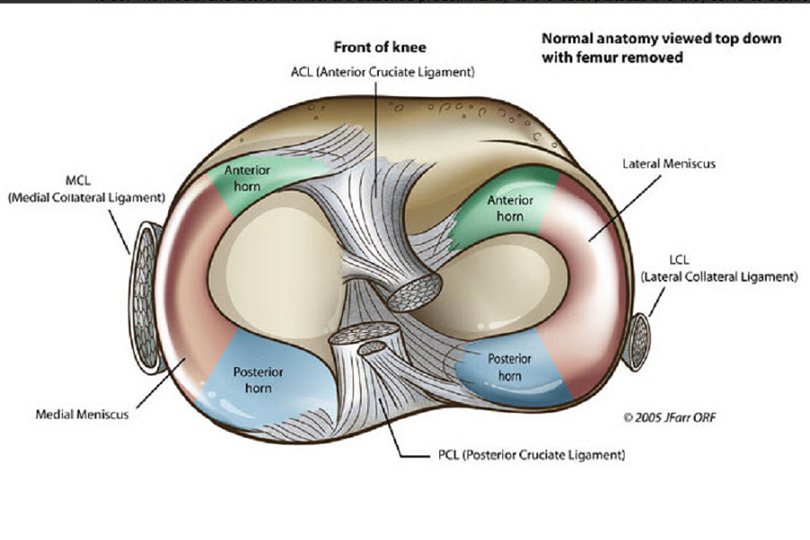 Question 1: What is the meniscus? What is its function? The meniscus is a C-shaped cartilage cushion that is positioned between the bones of the knee (the femur and tibia). It has several functions including acting as a shock absorber, a protector of the surface cartilage and a stabilizer of the knee. There are two menisci in each knee- a medial and lateral meniscus. The medial is positioned at the inside of the knee, the lateral to the outside. Both the medial and lateral meniscus increase the surface area of bone contact within the knee when walking/running, thereby spreading out the contact forces over a larger area. This function protects the surface gliding cartilage (called articular cartilage) from being damaged. Question 2: How is a meniscus torn/damaged? A meniscus can be torn in several ways, but most commonly it is damaged when the knee undergoes an abrupt change of direction and the stress is more than the meniscus can withstand. In the case of Castro, it sounds as though he had some damage to his meniscus in 2011 when he also tore his ACL. A meniscus tear accompanies an ACL tear about 50% of the time. Either the medial or lateral meniscus can be torn, and in some cases both sustain injury. Question 3: How are meniscus tears assessed? Most commonly an MRI is ordered to look at the meniscus more closely. The meniscus can’t be seen on xrays. There is a device that is similar to a large needle that has a camera inside it that can be inserted into the knee during an office visit to view the meniscus directly, but I don’t have much experience with this device. Question 4: What types of tears can occur? There are a number of different types of meniscus tears, and there are many tears which don’t fit neatly into a single category as well. Question 5: What are the treatment options for meniscus tears? There are a number of different options for treatment of meniscus tears. Not every meniscus tear will require surgery- in fact, many people may have a meniscus tear in their knee and not even be aware of it. Anti-inflammatory medication, injections (including cortisone, gel/lubricant, and PRP injections), and physical therapy are among the non-surgical options. When these are not successful (or surgery is deemed necessary early on), knee arthroscopy is performed. During knee arthroscopy, the knee is inflated with fluid and a camera is inserted inside the joint. The structures of the knee can be readily assessed, and tools are used to examine and probe the meniscus, surface cartilage and ligaments. The specifics of the meniscus tear are then evaluated. A decision is then made whether to repair the meniscus or remove the torn portion of meniscus (called a partial meniscectomy). This decision can be complex and is based on a number of factors including the size, location and orientation of the tear, patient-specific factors such as age, weight, health, prior surgery to the knee and others. Ideally, we would repair all meniscus tears, as removing meniscus can lead to the development of arthritis later on. However, some tears are simply unable to be repaired and must be partially removed. Tears closer to the outer edge of the meniscus have better blood supply are more likely to heal, and thus are better suited to repair. Question 6: What is the recovery time? This can vary significantly, but for partial removal, most athletes can return to sports between 6-10 weeks after surgery. For repair, the recovery is longer because of the additional time required for the repaired tissue to heal. Typically, a return to sports for a meniscus repair is around 3-5 months. Question 7: Are there any special circumstances with Castro’s knee? I would say yes. The fact that he has had two prior surgeries on the knee (including an ACL reconstruction) often makes the MRI more challenging to interpret and can complicate decision-making. It is unclear to me if the current meniscus damage is in the same area where he previously had surgery, which can create an already abnormal appearance on MRI. Thus, it is difficult to know whether what appears abnormal on a current MRI is ‘new’ or ‘old’. However, it sounds like he has continued to have intermittent problems over a lengthy amount of time now, and it seems reasonable to go ahead with arthroscopy. One concern is that a tear can enlarge in size over time if left alone (though this is difficult to predict). Dr. LaPrade is a world’s authority on knee problems and I’m sure he will do his best to get Castro back to action quickly and safely.
-
BREAKING NEWS: Jason Castro Out for the Year
Heezy1323 replied to Cody Christie's topic in Twins Daily Front Page News
This is definitely a bummer for Castro, but it may be best for the long-term health of his knee. For anyone who is interested, I wrote a blog post about meniscus injuries that some may find informative/helpful. http://twinsdaily.com/blog/1036/entry-11062-jason-castro-knee-surgery-qa/- 75 replies
-
- 9
-

-
- jason castro
- mitch garver
- (and 1 more)
-
Jason Castro Meniscus Surgery Q&A Heezy1323 Twins starting catcher Jason Castro went on the DL May 5th with pain in his right knee. Initially, it was reported that Castro underwent a cortisone injection and was hopeful to return to the lineup soon. Unfortunately, after traveling to Vail, CO to see noted orthopedic surgeon Dr. Robert LaPrade, Castro is now scheduled to undergo surgery May 15th to address a meniscus tear. (Update: Now being reported that he underwent more extensive surgery including a meniscus repair and is now out for the season). This will be the third surgery for Castro’s troublesome right knee, according to the Pioneer Press. He sustained an ACL tear along with a meniscus tear in spring training 2011 and underwent surgery, which caused him to miss the entire season. He also underwent arthroscopic surgery on the same knee in September 2013. Let’s discuss some knee anatomy and some meniscus ‘fun facts’ (Note: these facts may only be considered fun to me…) Question 1: What is the meniscus? What is its function? The meniscus is a C-shaped cartilage cushion that is positioned between the bones of the knee (the femur and tibia). It has several functions including acting as a shock absorber, a protector of the surface cartilage and a stabilizer of the knee. There are two menisci in each knee- a medial and lateral meniscus. The medial is positioned at the inside of the knee, the lateral to the outside. Both the medial and lateral meniscus increase the surface area of bone contact within the knee when walking/running, thereby spreading out the contact forces over a larger area. This function protects the surface gliding cartilage (called articular cartilage) from being damaged. Question 2: How is a meniscus torn/damaged? A meniscus can be torn in several ways, but most commonly it is damaged when the knee undergoes an abrupt change of direction and the stress is more than the meniscus can withstand. In the case of Castro, it sounds as though he had some damage to his meniscus in 2011 when he also tore his ACL. A meniscus tear accompanies an ACL tear about 50% of the time. Either the medial or lateral meniscus can be torn, and in some cases both sustain injury. Question 3: How are meniscus tears assessed? Most commonly an MRI is ordered to look at the meniscus more closely. The meniscus can’t be seen on xrays. There is a device that is similar to a large needle that has a camera inside it that can be inserted into the knee during an office visit to view the meniscus directly, but I don’t have much experience with this device. Question 4: What types of tears can occur? There are a number of different types of meniscus tears, and there are many tears which don’t fit neatly into a single category as well. Question 5: What are the treatment options for meniscus tears? There are a number of different options for treatment of meniscus tears. Not every meniscus tear will require surgery- in fact, many people may have a meniscus tear in their knee and not even be aware of it. Anti-inflammatory medication, injections (including cortisone, gel/lubricant, and PRP injections), and physical therapy are among the non-surgical options. When these are not successful (or surgery is deemed necessary early on), knee arthroscopy is performed. During knee arthroscopy, the knee is inflated with fluid and a camera is inserted inside the joint. The structures of the knee can be readily assessed, and tools are used to examine and probe the meniscus, surface cartilage and ligaments. The specifics of the meniscus tear are then evaluated. A decision is then made whether to repair the meniscus or remove the torn portion of meniscus (called a partial meniscectomy). This decision can be complex and is based on a number of factors including the size, location and orientation of the tear, patient-specific factors such as age, weight, health, prior surgery to the knee and others. Ideally, we would repair all meniscus tears, as removing meniscus can lead to the development of arthritis later on. However, some tears are simply unable to be repaired and must be partially removed. Tears closer to the outer edge of the meniscus have better blood supply are more likely to heal, and thus are better suited to repair. Question 6: What is the recovery time? This can vary significantly, but for partial removal, most athletes can return to sports between 6-10 weeks after surgery. For repair, the recovery is longer because of the additional time required for the repaired tissue to heal. Typically, a return to sports for a meniscus repair is around 3-5 months. Question 7: Are there any special circumstances with Castro’s knee? I would say yes. The fact that he has had two prior surgeries on the knee (including an ACL reconstruction) often makes the MRI more challenging to interpret and can complicate decision-making. It is unclear to me if the current meniscus damage is in the same area where he previously had surgery, which can create an already abnormal appearance on MRI. Thus, it is difficult to know whether what appears abnormal on a current MRI is ‘new’ or ‘old’. However, it sounds like he has continued to have intermittent problems over a lengthy amount of time now, and it seems reasonable to go ahead with arthroscopy. One concern is that a tear can enlarge in size over time if left alone (though this is difficult to predict). Dr. LaPrade is a world’s authority on knee problems and I’m sure he will do his best to get Castro back to action quickly and safely.
- 3 comments
-
- 3
-
-
- jason castro
- knee surgery
- (and 1 more)
-

From the album: Meniscus pics
-
-
-

From the album: Meniscus pics
-

From the album: Meniscus pics
-
Fair point. But I would say, in my experience at least, this is almost never the case. We typically try to give our best evaluation of the likelihood of success with both surgical and non-surgical treatment (and now sometimes including consideration of PRP and/or stem cell treatment). It is typically up to the player to decide which route they would like to take. Certainly in some cases, suggestions may be made from team/agent/family etc, but I have not experienced a case where a surgeon or team 'convinced' anyone they didn't need surgery. Doesn't mean it doesn't ever happen, but my thought would be it is quite unlikely. There are cases where the reverse is true, I would say- that sometimes an athlete thinks they can overcome an injury without surgery and a surgeon's experience tells them it's going to be very difficult. In which case I have had to strongly recommend surgery to certain patients. But, even then, it is their choice. All we can do is provide the best information we can to let them make the best choice for themselves.
-
Thanks for the compliment! I don't have a lot of experience writing blog posts such as this, so I welcome feedback about how I can be better.
-
Thanks! I am an orthopedic surgeon who specializes in sports medicine. I also was able to spend a year training with Dr. Andrews in Pensacola, so I have some insight into what goes on during these type of situations from my experience there as well.
-
News came down yesterday that highly-touted Twins SS prospect Wander Javier will undergo season-ending surgery on his left (non-throwing) shoulder to repair a torn labrum. According to LEN, this was an injury that was suffered initially late last season, but initially the decision was made to attempt to treat Javier with rehabilitation. Unfortunately, he has continued to have pain in the shoulder, and he has taken the dreaded trip to Pensacola, FL to see Dr. James Andrews. It sounds like Dr. Andrews will perform surgery soon, and that Javier is expected to be out six to nine months. Shoulder injuries are common in baseball players, certainly. But they are much more common in the throwing shoulder, particularly in pitchers. Let’s discuss how this injury may be similar and different. News came down yesterday that highly-touted Twins SS prospect Wander Javier will undergo season-ending surgery on his left (non-throwing) shoulder to repair a torn labrum. According to LEN, this was an injury that was suffered initially late last season, but initially the decision was made to attempt to treat Javier with rehabilitation. Unfortunately, he has continued to have pain in the shoulder, and he has taken the dreaded trip to Pensacola, FL to see Dr. James Andrews. It sounds like Dr. Andrews will perform surgery soon, and that Javier is expected to be out six to nine months. Shoulder injuries are common in baseball players, certainly. But they are much more common in the throwing shoulder, particularly in pitchers. Let’s discuss how this injury may be similar and different. Question 1: What is a labrum, anyway? The shoulder is considered a ball-and-socket joint. The round ball (humeral head) sits in the socket (glenoid) similar to how a golf ball sits on a golf tee. Around the perimeter of the golf tee is a strong cartilage tissue called a labrum. The labrum surrounds the socket similar to the red gasket on a mason jar lid. Its function is to help act as a ‘bumper’ to hold the golf ball on the golf tee. It is also an attachment point for ligaments around the shoulder that also contribute to shoulder stability. Question 2: How does a labrum get injured? There are several ways that someone could injure the labrum of the shoulder. In baseball players (and pitchers in particular), it is common for a labrum tear to occur at the top of the socket near the attachment of the biceps tendon. This is called a SLAP tear (Superior Labrum Anterior to Posterior) and is typically the cumulative result of repetitive use. However, it can also occur as a result of a dive/fall if the mechanism is just right. Another way the labrum can be injured is as a result of a shoulder dislocation. In the majority of cases, the ball dislocates out the front (anterior) of the socket, and the attachment of the labrum to the rim of the socket is damaged. A final way that I have seen labrum injuries in baseball players is in the front shoulder of hitters. Occasionally, as a result of an aggressive swing (or combination of swings) a player can damage the labrum in the back part (posterior) of the shoulder. This is also an injury that I sometimes see in golfers. In Javier’s case, the injury is to his left shoulder. As he is a right-handed hitter, this would be his front shoulder. I was not able to find any reports of him dislocating his shoulder last season (which would suggest an anterior labral tear). It is possible, then, that his labral tear is more in the back of the socket (though this is purely speculation on my part). Question 3: Why didn’t he just have surgery in the off-season to get this taken care of? This is always a difficult question to answer without knowing specifics of the situation. Hindsight is 20/20, but it is not always known at the time of an initial injury whether it is going to require surgery or whether rehab will be sufficient. Clearly, the Twins and Javier felt that rehab stood a reasonable chance of being successful, or I suspect surgery would have been undertaken previously. Question 4: What is done at surgery? There are subtle variations in technique for these type of injuries, but the majority are treated with arthroscopic surgery. This means a small fiber-optic camera is inserted into the shoulder, and the shoulder is filled with fluid. The labrum is then examined and the extent of the tear is assessed. Typically, the pre-op MRI will give the surgeon a good idea how extensive the tear is, but the precise size and location of the tear is not known until surgery. Once the tear has been assessed, the repair process begins. Small anchors (similar to plastic drywall screws) are inserted into the rim of the socket in the area of the labrum damage. These vary in size, but are typically somewhere around 3.0mm in diameter. These anchors have strong stitches attached. The stitches are passed around the labrum using special tools and the labrum is secured back to the rim of the socket. This process is repeated for as many anchors as are needed to completely repair the tear. After surgery, the small incisions are closed with suture and the patient heads to the recovery room. Surgery typically takes around 1-1.5 hours. Question 5: What happens after surgery? Why does it take so long to get back to full activity? Any time we repair a structure in orthopedics, the rate of healing is dependent on a number of factors. Some factors include the type of tissue injured, the severity of the injury, the age and health of the patient as well as numerous others. One helpful comparison in this case can be a fracture of a bone. Bone, as compared to cartilage (labrum) has a much greater blood supply. As such, most bone fractures are able to fully heal somewhere around 6-10 weeks (with some variability of course). A labrum (as with all cartilage) has a much poorer blood supply, and as a result takes a lot longer to heal. This is one of the main reasons for the lengthy rehab. In addition, the stresses placed on the shoulder are significant. The labrum has to withstand a tremendous amount of force when throwing a baseball or swinging a bat. The muscles around the shoulder tend to get weak quickly after surgery, and it takes time to rebuild the muscles. Proceeding too aggressively (before the muscles have recovered) places extra stress on the labrum, and can lead to failure of the repair to fully heal. Question 6: Will Javier’s shoulder ever be the same? This is perhaps the most difficult question to answer. Without knowing the extent and location of the damage, it is very difficult to comment on this subject. It is certainly better that this is his non-throwing shoulder, as that likely portends a better chance of full recovery. Few surgeons have done more of this type of surgery than Dr. Andrews, so he is in good hands. View full article
-
 Question 1: What is a labrum, anyway? The shoulder is considered a ball-and-socket joint. The round ball (humeral head) sits in the socket (glenoid) similar to how a golf ball sits on a golf tee. Around the perimeter of the golf tee is a strong cartilage tissue called a labrum. The labrum surrounds the socket similar to the red gasket on a mason jar lid. Its function is to help act as a ‘bumper’ to hold the golf ball on the golf tee. It is also an attachment point for ligaments around the shoulder that also contribute to shoulder stability. Question 2: How does a labrum get injured? There are several ways that someone could injure the labrum of the shoulder. In baseball players (and pitchers in particular), it is common for a labrum tear to occur at the top of the socket near the attachment of the biceps tendon. This is called a SLAP tear (Superior Labrum Anterior to Posterior) and is typically the cumulative result of repetitive use. However, it can also occur as a result of a dive/fall if the mechanism is just right. Another way the labrum can be injured is as a result of a shoulder dislocation. In the majority of cases, the ball dislocates out the front (anterior) of the socket, and the attachment of the labrum to the rim of the socket is damaged. A final way that I have seen labrum injuries in baseball players is in the front shoulder of hitters. Occasionally, as a result of an aggressive swing (or combination of swings) a player can damage the labrum in the back part (posterior) of the shoulder. This is also an injury that I sometimes see in golfers. In Javier’s case, the injury is to his left shoulder. As he is a right-handed hitter, this would be his front shoulder. I was not able to find any reports of him dislocating his shoulder last season (which would suggest an anterior labral tear). It is possible, then, that his labral tear is more in the back of the socket (though this is purely speculation on my part). Question 3: Why didn’t he just have surgery in the off-season to get this taken care of? This is always a difficult question to answer without knowing specifics of the situation. Hindsight is 20/20, but it is not always known at the time of an initial injury whether it is going to require surgery or whether rehab will be sufficient. Clearly, the Twins and Javier felt that rehab stood a reasonable chance of being successful, or I suspect surgery would have been undertaken previously. Question 4: What is done at surgery? There are subtle variations in technique for these type of injuries, but the majority are treated with arthroscopic surgery. This means a small fiber-optic camera is inserted into the shoulder, and the shoulder is filled with fluid. The labrum is then examined and the extent of the tear is assessed. Typically, the pre-op MRI will give the surgeon a good idea how extensive the tear is, but the precise size and location of the tear is not known until surgery. Once the tear has been assessed, the repair process begins. Small anchors (similar to plastic drywall screws) are inserted into the rim of the socket in the area of the labrum damage. These vary in size, but are typically somewhere around 3.0mm in diameter. These anchors have strong stitches attached. The stitches are passed around the labrum using special tools and the labrum is secured back to the rim of the socket. This process is repeated for as many anchors as are needed to completely repair the tear. After surgery, the small incisions are closed with suture and the patient heads to the recovery room. Surgery typically takes around 1-1.5 hours. Question 5: What happens after surgery? Why does it take so long to get back to full activity? Any time we repair a structure in orthopedics, the rate of healing is dependent on a number of factors. Some factors include the type of tissue injured, the severity of the injury, the age and health of the patient as well as numerous others. One helpful comparison in this case can be a fracture of a bone. Bone, as compared to cartilage (labrum) has a much greater blood supply. As such, most bone fractures are able to fully heal somewhere around 6-10 weeks (with some variability of course). A labrum (as with all cartilage) has a much poorer blood supply, and as a result takes a lot longer to heal. This is one of the main reasons for the lengthy rehab. In addition, the stresses placed on the shoulder are significant. The labrum has to withstand a tremendous amount of force when throwing a baseball or swinging a bat. The muscles around the shoulder tend to get weak quickly after surgery, and it takes time to rebuild the muscles. Proceeding too aggressively (before the muscles have recovered) places extra stress on the labrum, and can lead to failure of the repair to fully heal. Question 6: Will Javier’s shoulder ever be the same? This is perhaps the most difficult question to answer. Without knowing the extent and location of the damage, it is very difficult to comment on this subject. It is certainly better that this is his non-throwing shoulder, as that likely portends a better chance of full recovery. Few surgeons have done more of this type of surgery than Dr. Andrews, so he is in good hands.
-
Thanks everyone. My first crack at a blog post like this- so I'm certainly open to feedback with regards to how I can make them more informative and helpful for others. Feel free to respond with any critiques or advice, or even suggested topics for future blog posts.
-
I agree that this would have been the ideal situation. But I can tell you from experience that in many cases (perhaps the majority) the decision to opt for surgery is far from clear. And the return to play rate from labrum surgery in baseball is not 100% (though I suspect it is better for non-throwing shoulders than it is for throwing shoulders). It always seems like an easier call with the benefit of hindsight.
-
I wrote a blog post that discusses some of this topic. Please feel free to review if you'd like. Non-throwing shoulders are preferable (if you had to pick one), but still see significant stress when batting. http://twinsdaily.com/blog/1036/entry-11043-wander-javier-injury-qa/
-
News came down yesterday that highly-touted Twins SS prospect Wander Javier will undergo season-ending surgery on his left (non-throwing) shoulder to repair a torn labrum. According to LEN, this was an injury that was suffered initially late last season, but initially the decision was made to attempt to treat Javier with rehabilitation. Unfortunately, he has continued to have pain in the shoulder, and he has taken the dreaded trip to Pensacola, FL to see Dr. James Andrews. It sounds like Dr. Andrews will perform surgery soon, and that Javier is expected to be out six to nine months. Shoulder injuries are common in baseball players, certainly. But they are much more common in the throwing shoulder, particularly in pitchers. Let’s discuss how this injury may be similar and different. Question 1: What is a labrum, anyway? The shoulder is considered a ball-and-socket joint. The round ball (humeral head) sits in the socket (glenoid) similar to how a golf ball sits on a golf tee. Around the perimeter of the golf tee is a strong cartilage tissue called a labrum. The labrum surrounds the socket similar to the red gasket on a mason jar lid. Its function is to help act as a ‘bumper’ to hold the golf ball on the golf tee. It is also an attachment point for ligaments around the shoulder that also contribute to shoulder stability. Question 2: How does a labrum get injured? There are several ways that someone could injure the labrum of the shoulder. In baseball players (and pitchers in particular), it is common for a labrum tear to occur at the top of the socket near the attachment of the biceps tendon. This is called a SLAP tear (Superior Labrum Anterior to Posterior) and is typically the cumulative result of repetitive use. However, it can also occur as a result of a dive/fall if the mechanism is just right. Another way the labrum can be injured is as a result of a shoulder dislocation. In the majority of cases, the ball dislocates out the front (anterior) of the socket, and the attachment of the labrum to the rim of the socket is damaged. A final way that I have seen labrum injuries in baseball players is in the front shoulder of hitters. Occasionally, as a result of an aggressive swing (or combination of swings) a player can damage the labrum in the back part (posterior) of the shoulder. This is also an injury that I sometimes see in golfers. In Javier’s case, the injury is to his left shoulder. As he is a right-handed hitter, this would be his front shoulder. I was not able to find any reports of him dislocating his shoulder last season (which would suggest an anterior labral tear). It is possible, then, that his labral tear is more in the back of the socket (though this is purely speculation on my part). Question 3: Why didn’t he just have surgery in the off-season to get this taken care of? This is always a difficult question to answer without knowing specifics of the situation. Hindsight is 20/20, but it is not always known at the time of an initial injury whether it is going to require surgery or whether rehab will be sufficient. Clearly, the Twins and Javier felt that rehab stood a reasonable chance of being successful, or I suspect surgery would have been undertaken previously. Question 4: What is done at surgery? There are subtle variations in technique for these type of injuries, but the majority are treated with arthroscopic surgery. This means a small fiber-optic camera is inserted into the shoulder, and the shoulder is filled with fluid. The labrum is then examined and the extent of the tear is assessed. Typically, the pre-op MRI will give the surgeon a good idea how extensive the tear is, but the precise size and location of the tear is not known until surgery. Once the tear has been assessed, the repair process begins. Small anchors (similar to plastic drywall screws) are inserted into the rim of the socket in the area of the labrum damage. These vary in size, but are typically somewhere around 3.0mm in diameter. These anchors have strong stitches attached. The stitches are passed around the labrum using special tools and the labrum is secured back to the rim of the socket. This process is repeated for as many anchors as are needed to completely repair the tear. After surgery, the small incisions are closed with suture and the patient heads to the recovery room. Surgery typically takes around 1-1.5 hours. Question 5: What happens after surgery? Why does it take so long to get back to full activity? Any time we repair a structure in orthopedics, the rate of healing is dependent on a number of factors. Some factors include the type of tissue injured, the severity of the injury, the age and health of the patient as well as numerous others. One helpful comparison in this case can be a fracture of a bone. Bone, as compared to cartilage (labrum) has a much greater blood supply. As such, most bone fractures are able to fully heal somewhere around 6-10 weeks (with some variability of course). Labrum (as with all cartilage) has a much poorer blood supply, and as a result takes a lot longer to heal. This is one of the main reasons for the lengthy rehab. In addition, the stresses placed on the shoulder are significant. The labrum has to withstand a tremendous amount of force when throwing a baseball or swinging a bat. The muscles around the shoulder tend to get weak quickly after surgery, and it takes time to rebuild the muscles. Proceeding too aggressively (before the muscles have recovered) places extra stress on the labrum, and can lead to failure of the repair to fully heal. Question 6: Will Javier’s shoulder ever be the same? This is perhaps the most difficult question to answer. Without knowing the extent and location of the damage, it is very difficult to comment on this subject. It is certainly better that this is his non-throwing shoulder, as that likely portends a better chance of full recovery. Few surgeons have done more of this type of surgery than Dr. Andrews, so he is in good hands.
- 3 comments
-
- 5
-
-
- wander javier
- shoulder injury
- (and 2 more)
-
Also not an area of my expertise, but my understanding is that most non-vegetarians are able to obtain plenty of B12 in their diet without supplementation. Also, oral B12 is very minimally absorbed, so injection is a preferred method of administration for those who need it. Polanco's situation is sure disappointing. You'd like to think that professional athletes would be ultra-careful about what they are putting in their bodies, but it's hard for me to speculate on the dynamics of the relationship between Polanco and his trainer/trainers in the DR. Hopefully he can use this as motivation and a learning experience, and be better for it on his return. I also hope it will be cause for others in the Twins system to use greater caution with supplements in the future as well (though one would like to think that Santana's prior situation would have been instructive in that sense).
-
Sorry for the delayed response, guys/gals. I hadn't read this thread until just today. A couple thoughts I have with respect to this development, with a couple of disclaimers... Disclaimer 1: I'm not a hand surgeon (I specialize in orthopedic sports medicine, so I operate mostly on shoulders/knees/hips/elbows) so I don't have much personal experience with this type of surgery/recovery Disclaimer 2: I'm still not certain I understand PRECISELY what was done for Santana If anyone still cares to hear my opinions after the two above points... well, here goes. If I remember correctly, he was immobilized in a cast or splint of some type after surgery (I think I recall seeing that somewhere). This is typically done to protect repaired tissue by immobilizing the surrounding joints. The plus side is that the tissue staying quiet/still allows the healing to progress without the intermittent micro trauma that can occur with movement. The down side is that keeping any joint immobilized for a prolonged period of time can lead to stiffness. Some joints are more prone to becoming stiff than others, and in my experience, elbows and the joints of the fingers are among those joints. So the duration of the immobilization is determined by trying to find that 'sweet spot' between protecting the tissue long enough that healing can occur relatively unimpeded, but not so long that significant stiffness begins to set in. To complicate things further, not everyone becomes stiff at the same rate, so in most cases, this is an educated guess. As such, being immobilized for several weeks after surgery on your MCP joint (as in Santana's case) is essentially guaranteed to impart some level of stiffness. Hopefully it is overcome relatively quickly with appropriate therapy and stretching after removal of the cast/splint. The issues to overcome include the scar tissue forming at the joint where surgery was performed and also stiffness of the tendon gliding in the tunnel on the palm side of the finger that helps pull the finger down. With respect to timeline, it's really hard for me to say given my relative inexperience in this area. To me, the level of concern regarding his stiffness is pretty low. It is fairly likely (to me) that he recovers his range of motion pretty quickly. To me the larger concern is going to be how his finger responds when he begins throwing. Is he going to have resolution of the symptoms he was experiencing that necessitated the surgery? Is he going to have 'flare-ups' that prolong the recovery? Are his pitches going to have the same control and 'bite'? Is he going to have to change the way he grips or throws any pitches due to this? Without examining him or having more information, it's hard to make any firm statements. My personal opinion would be that going from not being able to hold a baseball to pitching in a major league game in 6 weeks seems... aggressive. I sure hope I'm wrong or being conservative. I'd love to see him on the bump sooner than later.


